
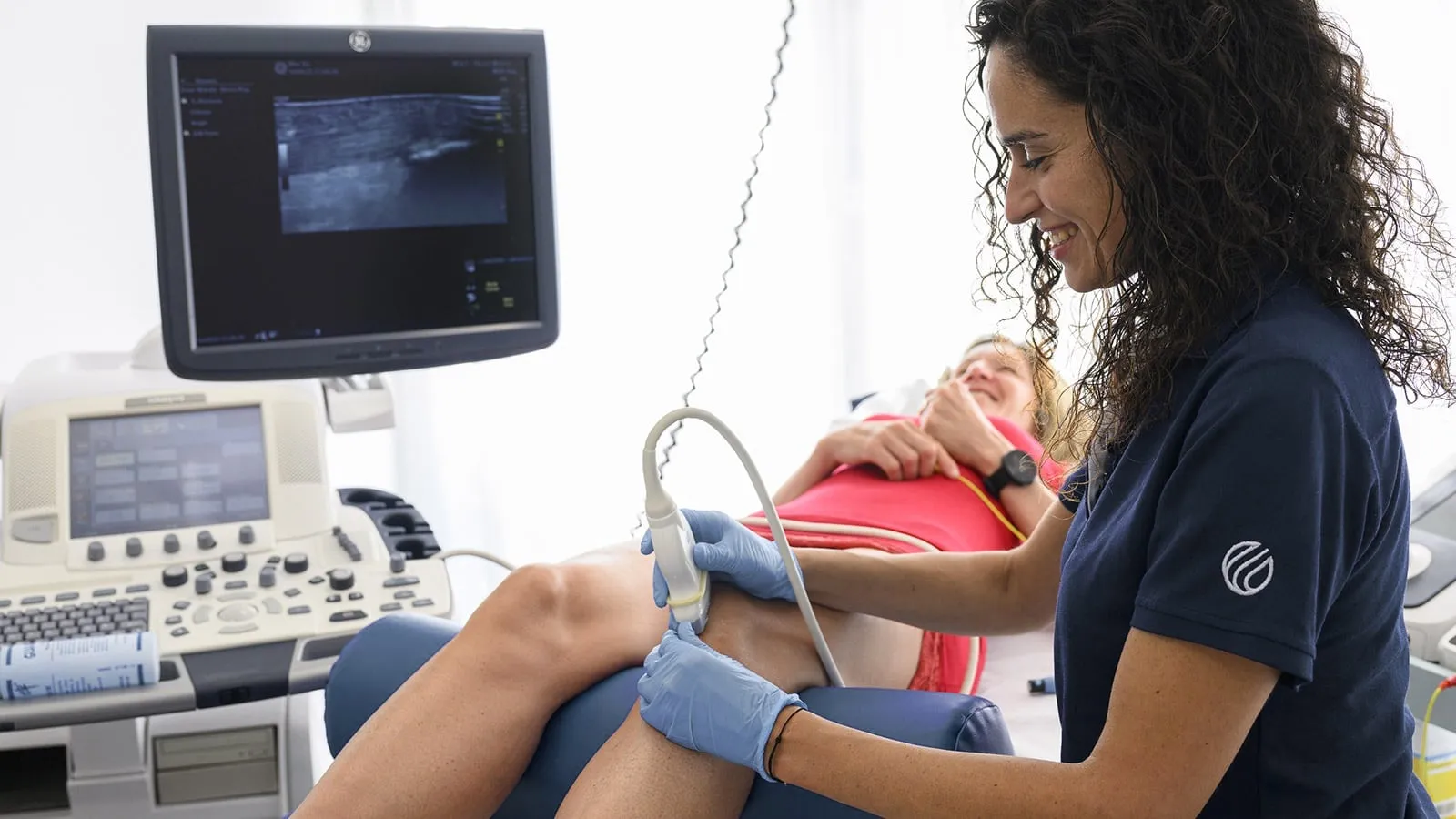
Percutaneous musculoskeletal electrolysis (EPM) is part of invasive physiotherapy. Under ultrasound, the therapist guides an acupuncture needle into the damaged tissue and applies a galvanic current there. The current causes an acute, local and controlled inflammation: the body clears the damaged tissue and repairs the soft tissue.
Compared with the EPTE technique, which uses low-voltage currents, EPM works with a higher current intensity. EPM protocols are based on clinical and laboratory studies.
Treatment takes place once a week and lasts 45 to 60 minutes. The first effects are often noticed after one or two sessions.
Other physiotherapeutic forms are used as a complement to this technique, such as muscle strengthening and stimulation of neuromuscular control through active exercises and other conventional physiotherapeutic treatments (myofascial treatments, diathermy…).
This technique is practiced by a therapist specialized in musculoskeletal invasive physiotherapy (MIP), who, in order to practice it, must first take a course in musculoskeletal ultrasound (minimum 30 hours) and then the EPM course (60 hours). This training is also included in a university master’s course that takes place in Spain, at San Pablo CEU University in Madrid (60 ECTS). During the training all the techniques of invasive physiotherapy are learned, which aim to treat soft tissues such as tendons, muscles, capsules, bursae, nerves and ligaments.
The technique is relatively new: it has been applied in Spain since 2008. The inventor of the technique is Jose Manuel Sánchez-Ibáñez, who registered the trademark EPI®. Later, Francisco Minaya-Muñoz and Fermín Valera-Garrido developed the EPM application methodology, which today is the most scientifically validated approach. Francisco Minaya-Muñoz and Fermín Valera-Garrido formed the MVClinic, which, in collaboration with San Pablo CEU University in Madrid, developed protocols for the treatment of each individual structure based on clinical and laboratory studies. In ten years, they trained many Spanish physiotherapists.
Indications
- Chronic tendinopathies (patellar tendon, Achilles tendon, pubalgia, lateral/medial epicondylitis, rotator cuff, supraspinatus tendon…)
- Bursitis (retrocalcaneal, deltoid bursitis, Achilles bursitis…)
- Acute and chronic muscle injuries
- Peripheral nerve entrapment syndromes
- Joint impingements
- Tenosynovitis (De Quervain’s syndrome, tenosynovitis of tibialis posterior…)
- Tibial periostitis
- Plantar fasciitis
- Acute and chronic muscle rupture (fibrosis)
- Myofascial trigger points
- Ligament sprain (medial collateral ligament of the knee, anterior talofibular ligament of the ankle…)
- Knee flexion contracture (flexum) (posterior joint capsule)
- Carpal and tarsal tunnel syndrome
- Anterolateral and posterior ankle impingement
- Adherent and painful scars
Advantages
- It is a precise and local treatment that, with the help of an ultrasound, is performed directly on the altered and/or degenerated tissue. The precision of this technique is also ensured through the use of Power Doppler and elastography.
- When the repair of connective tissue in a tendon or muscle is difficult, the EPM technique starts a new process of collagen fibre proliferation, which in turn allows the repair process to resume physiologically.
- The structural changes and mechano-biological behavior of soft tissue are immediate and in real time.
- The efficiency of EPM is high compared with both conventional physiotherapy (ultrasound, laser, Cyriax technique, diathermy therapy, shock waves, etc.) and other medical treatments (infiltrations or sometimes even surgery) in cases of neuro-musculoskeletal problems.
- The frequency of relapse is low.
In chronic cases
In more chronic cases, electrolysis can be combined with other invasive physiotherapy techniques:
- Neuromodulation (NMP_e)
- Microcurrents
- Bioregulation
- Intramuscular modulation (IMS)
- High-voltage currents
Contraindications
- Belonephobia
- Acute systemic infections with or without fever
- No clear consent from the patient
- All cases of acute emergency
- Osteosynthesis, prostheses, and implants (local contraindication)
- Pacemakers
- Heart disease where electric field changes could alter heart rhythm
- Skin problems (local contraindication)
- Thrombophlebitis
- Tumors
- Sensitivity disorders (relative contraindication)
- Lymphoedema or removal of lymph nodes (local contraindication)
- High risk of infection (relative contraindication)
- Metabolic alterations (relative contraindication)
- Fibromyalgia (relative contraindication)
- Pregnancy (absolute contraindication in the first trimester and in the subsequent months relative to areas that could affect the fetus)
- Children (parental permission required)
- Contagious diseases (relative contraindication)
- Allergies to the material used (relative contraindication, special measures required)
- Epilepsy (relative contraindication)
- Mucous membranes, eyes, genitals, nipples, and endocrine glands
Studies
Although the technique is relatively new, several studies have already assessed its effect:
- Galvanic current activates the NLRP3 inflammasome to promote Type I collagen production in tendon
- A Molecular Mechanisms of Regeneration in Chronic Tendinopathy Using Ultrasound-Guided Intratissue Percutaneous Electrolysis (EPI®)
- Ultrasound-guided percutaneous needle electrolysis in chronic lateral epicondylitis: short-term and long-term results
- Ultrasound-Guided Percutaneous Needle Electrolysis and Rehab and Reconditioning Program for Rectus Femoris Muscle Injuries: A Cohort Study with Professional Soccer Players and a 20-Week Follow-Up
- Randomized controlled trial comparing the effectiveness of the ultrasound-guided galvanic electrolysis technique (USGET) versus conventional electro-physiotherapeutic treatment on patellar tendinopathy
- Intratissue Percutaneous Electrolysis vs Corticosteroid Infiltration for the Treatment of Plantar Fasciosis
- Treatment of proximal hamstring tendinopathy-related sciatic nerve entrapment: presentation of an ultrasound-guided “Intratissue Percutaneous Electrolysis” application
- The Effectiveness of Minimally Invasive Techniques in the Treatment of Patellar Tendinopathy: A Systematic Review and Meta-Analysis of Randomized Controlled Trials
- Ultrasound-Guided Percutaneous Needle Electrolysis in Dancers with Chronic Soleus Injury: A Randomized Clinical Trial
- Percutaneous Needle Electrolysis Accelerates Functional Muscle Regeneration in Mice
Studies conducted in Spain have shown that damaged tissue can potentially be treated through the EPM procedure rather than surgery. This implies that the affected tendon or area is able to heal without the need for invasive surgery.